When you click on links to various merchants on this site and make a purchase, this can result in this site earning a commission. Affiliate programs and affiliations include, but are not limited to, the eBay Partner Network.
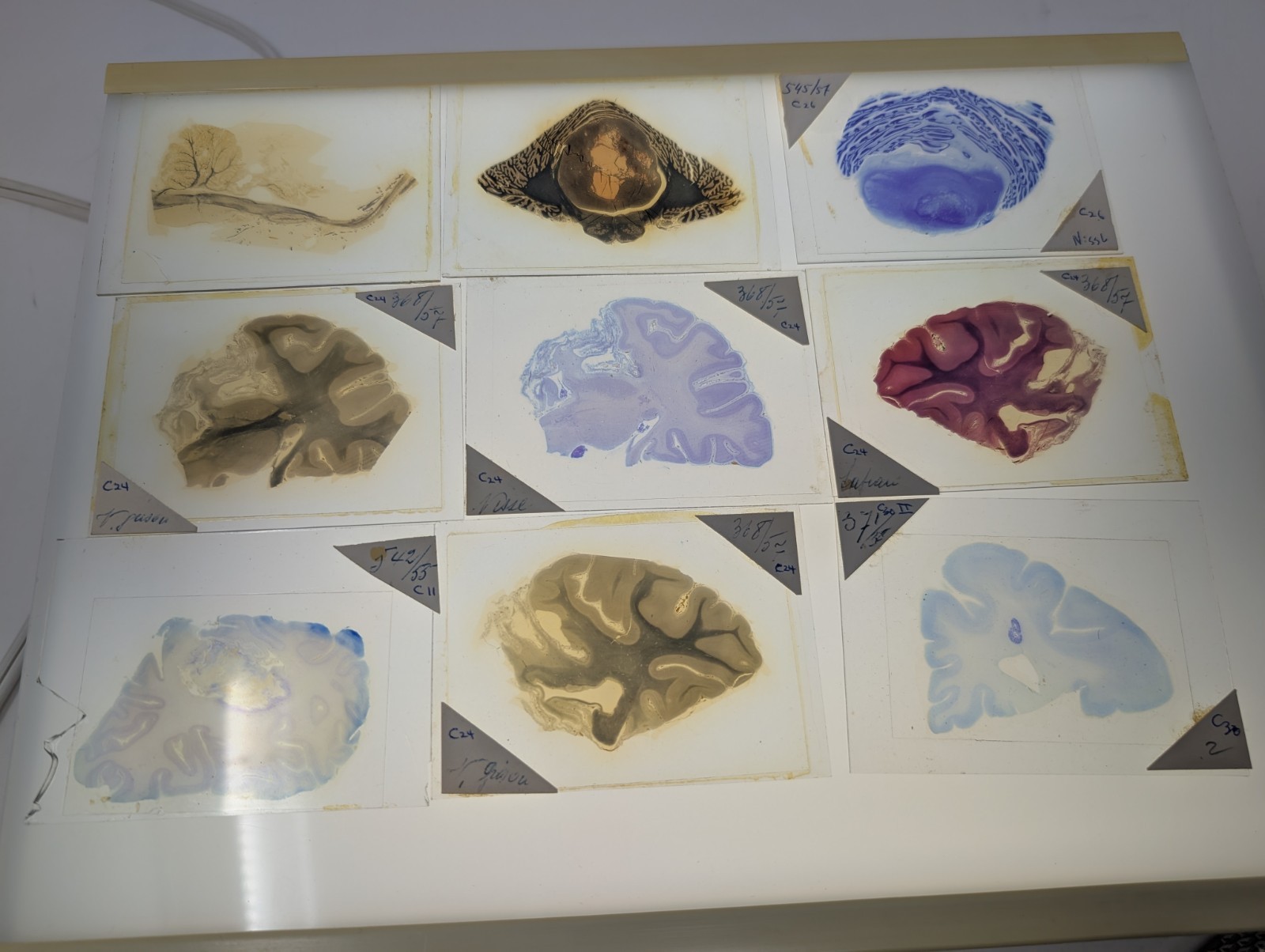
Rare vintage collection of histology microscope slides featuring stained cross-sections of mammalian brain and nervous system tissue mounted on glass.
The slides appear to include multiple regions of the brain such as the cerebrum, cerebellum, cortical folds, white matter tracts, and possible spinal cord or brainstem sections, with several handwritten anatomical labels visible.
The vivid staining methods highlight different neural structures and cell densities, creating striking abstract organic imagery in blue, violet, pink, sepia, and black tones.
Some slides show highly folded cortical tissue, while others display layered neural architecture and cross-sectional nerve structures typical of teaching or laboratory specimens used in medical, biological, or anatomical study.
Approx 3.5"x2.5"
Please view photos carefully for condition details